Cutler-Beard lid-sharing flap is a skin muscle-conjunctival flap that is extracted from the lower eyelid and progressed upon to make up for defects or flaws in the upper eyelid reaching up to 100% in width.
In several cases where complete upper lid sacrifice cannot be evaded and is imperative, a bridged advancement flap (as seen in the Cutler-Beard method) has been documented/shown to yield better, more appreciated results. The Cutler-Beard procedure was brought forth as a potential surgical approach to be adopted in the repair of the upper lid; it is premised upon the concept of sharing lower lids. Perhaps the most noticeable flaw from this technique is seen in its incapacity to sufficiently transfer tarsus to the upper lid and as a consequence, producing a floppy lid. A wide variety of grafts such as donor sclera, ear cartilage, and skin were used to repair the tarsus. Others such as the buccal cartilage, nasal septum, or even the tarsus from the opposing eye may also be employed in this procedure.
The Cutler-Beard treatment technique which has the replacement of upper eyelid tarsus with Achilles tendon “as an allograft” as an integral part of the process, yielded positive results and showed success in the treatment of major anomalies/issues in the upper eyelid. The human Achilles tendon is composed of collagen fibril bundles, which are configured and set up in parallel forms, along with supportive cells in dense connective tissue. It has been shown to possess measurable elasticity, viscosity, and plasticity and a counter to functional demands.
Looking at it from a general viewpoint; a great degree of safety and cost-effectiveness is attached to this procedure; and this is of course backed up by satisfactory cosmetic outcomes, pre- and post- operation.
The surgical repair of significant full-thickness defects of the upper eyelid caused by the resection of eyelid cancer is something of a surgical encumbrance. This procedure was initially proposed as an eyelid-sharing approach with two stages, to repair abnormalities involving more than half the total width of the upper eyelid. This approach entails the progression of a composite full-thickness lower eyelid flap, in a posterior position to the residual margin of the lower eyelid and then, into the upper eyelid defect. During the second part of the treatment; this happens between 6 – 8 weeks after the starting phase of the surgical process, the flap is lysed. The primary objective of this protocol is to restore the anatomy of the eyelid, with function and aesthetics appreciably preserved.
Not taking away from the fact that this operation has its own merits, it is worthy to note that the patient should be advised that the surgery is difficult and complex. Also, that the outcome will never act or look exactly like a normal lid, and that in given situations, the need for touch-up surgeries might arise. This is due to the fact that the lid is often thick and immutable to some extent after undergoing repair. The stability of the upper eyelid can be enhanced and built on with a tarsoconjunctival graft harvested from the upper eyelid on the side that was not affected by the defect.
At the end of your appointment with the surgeon, the need to have a Cutler-Beard flap surgery must have been established. The next line of action you will be taking will be preparing for the operation proper. The instructions the surgeon provided you with must be strictly adhered to – as this would go a long way in ensuring a successful operation and even speedy recovery thereafter. For instance, the use of medications with blood-thinning tendencies should be halted for quite a number of days provided that it is feasible.
The anesthesiologist is called upon to administer local anaesthesia to the patient, and the surgeon proceeds with the operation once the effect [of the anaesthesia] has set in. Forceps are then used to draw the medial and lateral upper lids toward one another, and a caliper is used in gauging the residual defect in a horizontal orientation. Then, the tarsal conjunctiva is noted and the eversion of contralateral upper the eyelid is achieved with the aid of a Desmarres retractor, ensuring that a 4mm surgical width is preserved from the eyelid's edges. This is followed by the harvesting of a tarsoconjunctival graft with the surgical blade being used to create the first incision involving both the conjunctiva and tarsus and the graft are eventually removed with a pair of Westcott scissors. Direct pressure is used to induce hemostasis, and the site where the graft was harvested from is left to heal on its own.
A line that is then made to the ipsilateral lower eyelid edge – with a 5mm margin preserved below it – is marked to gain surgical width. The surgeon then uses a special surgical blade to make an incision at the surgical spot, and thereafter uses Westcott scissors to complete a full-thickness incision along the lower eyelid. After this, a Jaeger lid plate is set in place to protect the globe. The inferior conjunctival fornix of the posterior lamella, as well as the anterior lamella's orbital rim, are then incised similarly – medially and laterally in an inferior direction. The harvested tarsoconjunctival graft is next placed with the surface of the conjunctiva towards the globe. This graft is eventually attached to the upper eyelid's remnant lateral and medial tarsal plates with absorbable sutures – the surgeon has to ensure proper alignment before doing this suturing. Again, another absorbable suture is then used to bind the graft's superior border to the upper eyelid conjunctiva edge revealing the cut. It is expedient that every conjunctival suture is applied with caution in this and future steps, with all knots facing anteriorly to avoid keratopathy and possible exposure of the suture to the globe. Thereafter, the surgeon firmly works the flap is onto the site of the upper eyelid defect. The surgeon will then dissect the conjunctiva from the flap's lower eyelid retractor and is then to the inferior border of the attached tarsoconjunctival graft and secured with a running absorbable suture. Another running absorbable suture is also used in binding the lower eyelid retractors and flap's orbicularis complex to the levator aponeurosis' cut edge. The flap's skin is next fastened to the upper eyelid skin using a combination of interrupted and running silk sutures if the aponeurosis has been entirely removed; otherwise, the sutures should secure the levator palpebrae superioris muscle.
Antibiotic eye ointment and a pressure eye patch are applied to all surgical sites. The lysis of the flap is the second part of the procedure, which is normally performed between 6 – 8 weeks after the initial procedure. In performing this, the patient is administered local anaesthesia. The surgeon will then insert a grooved director into the flap to prevent globe damage. A special surgical tool is now used in cutting the flap skin, even as a pair of Westcott scissors is used in completing the process at some safe distance from the point of the new upper eyelid margin. This goes a way in preventing keratopathy and the retraction of the eyelids after surgery.
To construct an anteriorly positioned mucocutaneous connection, the upper lid conjunctiva is moved anteriorly for about 2 mm onto the upper eyelid margin that is being created anew – using an absorbable running plain gut suture. By this, the risk of the patient suffering keratopathy as a result of keratinized skin is considerably minimized. Excess superior flap tissue is carefully removed in the event whereby the flap has been stretched since the initial step; this permits it to be firmly secured to the bridge of the lower eyelid. The granulation tissue of the bridge of the lower eyelid bridge is denuded with a surgical blade and the edge of the flap at the superior aspect is then joined to the bridge in a two-layered fashion using a combination of interrupted and running sutures. All surgical areas are treated with antibiotic ophthalmic ointment.
The surgeon will give you some medications and instructions directed at aiding your recovery. Hence, you should not fail to adhere to this. More so, a follow-up plan will be drawn up to enable close monitoring of your situation as you recover.
A procedure as complicated as Cutler-Beard flap needs to be performed under the supervision of the best team of plastic surgeons, with years of experience in the field. This is guaranteed at The Esthetic Clinics where Dr. Debraj Shome – India’s celebrity oculoplastic surgeon with many notable mentions and awards across the world – leads a vibrant group of cosmetic surgeons and plastic surgeons. More so, our medical facility is very ready to ensure the success of your Cutler-Beard flap surgery – we will see you through from the pre-operative phase to the postoperative aspect.
Concerning relating the cost of Cutler-Beard flap surgery; it is crucial that we have a first-hand evaluation of your condition to enable us with you with a workable estimate. That said, the Cutler-Beard flap procedure price will be dependent on the size of the defect, diagnosis, surgeon fee, prescriptions, and length of the follow-up period.
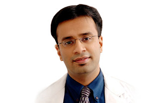

Dr. Debraj Shome is Director and Co founder of The Esthetic Clinics. He has been rated amongst the top surgeons in India by multiple agencies. The Esthetic Clinics patients include many international and national celebrities who prefer to opt for facial cosmetic surgery and facial plastic surgery in Mumbai because The Esthetic Clinics has its headquarters there.
